Home
Uncategories
Shoulder Muscles Diagram : Deltoid Muscle Wikipedia / The shoulder muscles bridge the transitions from the torso into the head/neck area and into the uppe.
Shoulder Muscles Diagram : Deltoid Muscle Wikipedia / The shoulder muscles bridge the transitions from the torso into the head/neck area and into the uppe.
Shoulder Muscles Diagram : Deltoid Muscle Wikipedia / The shoulder muscles bridge the transitions from the torso into the head/neck area and into the uppe.. The muscles in the shoulder aid in a wide range of movement and help protect and maintain the main shoulder joint, known as the glenohumeral joint. Neck muscle anatomy mri 12 photos of the neck muscle anatomy mri neck muscle anatomy images, neck muscle anatomy pictures, neck muscle anatomy posterior, neck muscle anatomy ultrasound, neck muscles anatomy radiology, human muscles, neck muscle anatomy images, neck muscle anatomy pictures, neck muscle anatomy. This flexibility is also what makes the shoulder prone to instability and injury. The bursa is a small sac of fluid that cushions and. The main shoulder muscles are trapezius, deltoid, pectoralis major and 4 rotator cuff muscles:
Related posts of diagram of shoulder muscles and tendons neck muscle anatomy mri. Plus, exercises for training them. The shoulder joint is formed where the humerus (upper arm bone) fits into the scapula (shoulder blade), like a ball and socket. Find out in this anatomy of the shoulder quiz. The shoulder blade (scapula) connects to the collarbone (clavicle) at this joint.
The Shoulder Joint Structure Movements And Muscles from www.martinpetkov.com The shoulder muscles are responsible for maintaining the widest range of motion of any joint in your body. It is a flat, gliding joint. The most common shoulder injuries are sprains, strains, and tears. This flexibility is also what makes the shoulder prone to instability and injury. Head , neck, shoulder, & back muscles— presentation transcript 2 muscles of the head and neck scalp. The tendons are the attachment of the muscle to the bone. These are located in the shoulder blade area, and each related tendon also attaches to the humerus. Find out in this anatomy of the shoulder quiz.
The human shoulder is made up of three.
The rotator cuff is a group of four muscles and tendons that surround the glenohumeral joint. It is a flat, gliding joint. The shoulder muscles are associated with movements of the upper limb shoulder muscles diagram. The most common shoulder injuries are sprains, strains, and tears. Shoulder muscles move the shoulder blades and upper arm bones. Related posts of diagram of shoulder muscles and tendons neck muscle anatomy mri. A muscle contracts to move bones; Each muscle of the shoulder assists with specific. The shoulder muscles and shoulder tendons involved with shoulder mobility include the four rotator cuff muscle and tendon pairs: The muscle elevates, depresses, rotates, and retracts the scapula, or shoulder blade. What are common rotator cuff injuries? The rotator cuff muscles and tendons may be injured by trauma, such as falling when skiing or biking, or from arthritic spurs that form within the shoulder and erode the cuff tissue over time. The shoulder joint is formed where the humerus (upper arm bone) fits into the scapula (shoulder blade), like a ball and socket.
Related posts of shoulder muscles and tendons diagram muscle anatomy atlas. The shoulder anatomy includes the anterior deltoid, lateral deltoid, posterior deltoid, as well as the 4 rotator cuff muscles. The rotator cuff is a group of four muscles and tendons that surround the glenohumeral joint. Or a broken collarbone most often happens from falling and landing directly onto your shoulder, such as while bicycling, skiing, or. Shoulder muscles and shoulder tendons.
Shoulder Anatomy Illustrations Healthy Shoulder Anatomy Shoulder Replacement Illustrations from www.medical-artist.com And the ligaments, which connect bones. Muscles allow us to move by pulling on bones. While seated, have your partner place one hand at the front of your shoulder joint and one hand at the rear. Shoulder muscles move the shoulder blades and upper arm bones. Head , neck, shoulder, & back muscles— presentation transcript 2 muscles of the head and neck scalp. The rotator cuff is a group of four muscles and tendons that surround the glenohumeral joint. Parts of the right shoulder blade: The muscles of the shoulder support and produce the movements of the shoulder girdle.they attach the appendicular skeleton of the upper limb to the axial skeleton of the trunk.
The muscles of the shoulder bridge the transitions from the torso into the head/neck area and into the upper extremities of the arms and hands.
Around the shoulder, muscles in the back, neck, shoulder, chest and upper arm all work together to support and move the shoulder. The bursa is a small sac of fluid that cushions and. The shoulder blade (scapula) connects to the collarbone (clavicle) at this joint. Muscles of the shoulder region. The acromioclavicular joint is formed by an articulation between the lateral end of the clavicle and the acromion process of the scapula. Shoulder mri radiographical and illustrated anatomical atlas from www.imaios.com a muscle contracts to move bones; The shoulder's flexibility can make it prone to injury. Parts of the right shoulder blade: Diagram of the shoulder, including the location of the rotator cuff. The main shoulder muscles are trapezius, deltoid, pectoralis major and 4 rotator cuff muscles: The shoulder anatomy includes the anterior deltoid, lateral deltoid, posterior deltoid, as well as the 4 rotator cuff muscles. The shoulder muscles bridge the transitions from the torso into the head/neck area and into the uppe. There are 10 muscles and 11 shoulder tendons related to shoulder mobility.
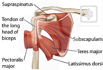
The shoulder muscles consist of the deltoids and the rotator cuff group.the deltoids are the muscles that can be seen on the outside of the body, whilst the rotator cuff group is found within the shoulder joint itself, providing structural support and allowing the shoulder to perform many functions. The supraspinatus, the infraspinatus, the teres minor and the subscapularis. Subscapularis, supraspinatus, infraspinatus and teres minor. To further reinforce the shoulder, the four muscles of the rotator cuff extend from the scapula and surround the head of the humerus to both rotate the arm and prevent dislocation. Related posts of diagram of shoulder muscles and tendons neck muscle anatomy mri.
Shoulder Tendons Shoulderdoc from www.shoulderdoc.co.uk The rotator cuff is a group of four muscles and tendons that surround the glenohumeral joint. Muscles allow us to move by pulling on bones. The rotator cuff muscles are important stabilizers and movers of the shoulder joint. The bursa is a small sac of fluid that cushions and. Diagram of the shoulder, including the location of the rotator cuff. The most common shoulder injuries are sprains, strains, and tears. The muscle elevates, depresses, rotates, and retracts the scapula, or shoulder blade. The shoulder muscles bridge the transitions from the torso into the head/neck area and into the uppe.
Each muscle of the shoulder assists with specific.
The rotator cuff muscles and tendons may be injured by trauma, such as falling when skiing or biking, or from arthritic spurs that form within the shoulder and erode the cuff tissue over time. The muscles in the shoulder aid in a wide range of movement and help protect and maintain the main shoulder joint, known as the glenohumeral joint. Muscles allow us to move by pulling on bones. The human shoulder is made up of three bones: Four of them are found on the anterior aspect of the shoulder, whereas the rest are located on the shoulder's posterior aspect and in the back. The tendons, which anchor muscle to bone; The main shoulder muscles are trapezius, deltoid, pectoralis major and 4 rotator cuff muscles: Muscle anatomy atlas 12 photos of the muscle anatomy atlas , human muscles. Shoulder mri radiographical and illustrated anatomical atlas from www.imaios.com a muscle contracts to move bones; The shoulder muscles consist of the deltoids and the rotator cuff group.the deltoids are the muscles that can be seen on the outside of the body, whilst the rotator cuff group is found within the shoulder joint itself, providing structural support and allowing the shoulder to perform many functions. Diagram of the shoulder, including the location of the rotator cuff. The muscle elevates, depresses, rotates, and retracts the scapula, or shoulder blade. Image via lh4.googleusercontent.com you can see in the shoulder muscle diagrams that the shoulder is one of the largest and most complex joints in the body.


0 Comments:
Posting Komentar